Human Back Bones Diagram - Human Bone Structure Back Human Back Bones Anatomy Human Anatomy Diagram Human Bones Anatomy Human Skeleton Anatomy Anatomy Bones - Collectively this region is called the vulva.
byAdmin-
0
Human Back Bones Diagram - Human Bone Structure Back Human Back Bones Anatomy Human Anatomy Diagram Human Bones Anatomy Human Skeleton Anatomy Anatomy Bones - Collectively this region is called the vulva.. This article looks at the anatomy of the back, including bones, muscles, and nerves. The human back extends from the buttocks to the posterior portion of the neck and shoulders.it is opposite from the chest, and the vertebral column runs down the back. Cervical bones diagram 12 photos of the cervical bones diagram cervical bones diagram, cervix anatomy diagram related posts of human back bones diagram human bone parts name. Bone structure diagram human foot. Each region has a number of vertebral bones.
Money back guarantee refund in 15 days. Related posts of female body back side anatomy skeleton bones diagram. The first seven bones (vertebrae) of your spine form your neck. Cervical bones diagram 12 photos of the cervical bones diagram cervical bones diagram, cervix anatomy diagram related posts of human back bones diagram human bone parts name. Just need a glimpse, leave your valuable advice let us know , and subscribe us!
Labeled Human Anatomy Diagram Of Man S Arm Shoulder And Upper Back Stock Images Page Everypixel from media.istockphoto.com Human backbone diagram, bone, human backbone diagram. They help support particular bones and make them move. Atlas (c1) the atlas is the first cervical vertebra and therefore abbreviated c1. Using this atlas of human anatomy of the spine and back. For more anatomy content please follow us and visit our website: Muscles of the abdomen lower back and pelvis. This article looks at the anatomy of the back, including bones, muscles, and nerves. Its appearance is different from the other spinal vertebrae.
Female reproductive organs of the lower torso.
Related posts of human back bones diagram pelvic bone labeled. Bones of the pelvis and lower back. They are the bones of your forearm. For more anatomy content please follow us and visit our website: Flat bones follow the process of intramembranous ossification where the young bones grow from a primary ossification center in fibrous membranes and leave a small region of. The spine diagram the spine diagram shown below, consists of many bones or vertebrae,soft discs,the spinal cord, and spinal nerves. Human backbone diagram, bone, human backbone diagram. Pelvic bone labeled 12 photos of the pelvic bone labeled pelvic bone labeled, pelvic bone labeling quiz, pelvic bone with labeling, pelvic girdle bone labeling quiz, pubic bone labeled, bone, pelvic bone labeled, pelvic bone labeling quiz, pelvic bone with labeling, pelvic girdle bone labeling quiz, pubic bone labeled As a person ages, these bones grow together and fuse into larger bones, leaving adults with only 206 bones. Collectively this region is called the vulva. Each region has a number of vertebral bones. At birth, the skeleton of a newborn has more than 300 bones; Related posts of human anatomy female lower back muscle anatomy triceps.
Human back bone chart back bones diagram human anatomy. As a person ages, these bones grow together and fuse into larger bones, leaving adults with only 206 bones. Vertebrae separated by intervertebral discs. Carpals—8 small bones of the wrist — includes the scaphoid, lunate, capitate, trapezium, and others. Collectively this region is called the vulva.
Male Anatomy From The Back Human Body Organs Anatomy Organs Human Anatomy Female from i.pinimg.com See lumbar spine anatomy diagram stock video clips. Immune and lymphatic systems of the lower torso. The spine diagram the spine diagram shown below, consists of many bones or vertebrae,soft discs,the spinal cord, and spinal nerves. Related posts of human back bones diagram pelvic bone labeled. They help support particular bones and make them move. Female reproductive organs of the lower torso. The spinal cord begins at the base of the brain and extends into the pelvis. This vertebra supports the skull.
The atlas is a ring of bone made up of two lateral masses joined at.
Collectively this region is called the vulva. Related posts of human anatomy female lower back muscle anatomy triceps. Just need a glimpse, leave your valuable advice let us know , and subscribe us! Our latest youtube film is ready to run. This article looks at the anatomy of the back, including bones, muscles, and nerves. Human spine diagram reading industrial wiring diagrams. This shopping feature will continue to load items when the enter key is pressed. The vertebrae, which stack like spools of thread, support the back and protect the spinal cord. Immune and lymphatic systems of the lower torso. Many muscles that move the trunk and legs, such as our abdominal muscles, attach to the hip bones. Bones, discs, and joints in your lower back your lower back contains 5 vertebral bones stacked above each other with intervertebral discs in between. Cheek bone (zygoma) upper jaw (maxilla). The smallest bone in the human body is called the stirrup bone, located deep inside the ear.
The human back extends from the buttocks to the posterior portion of the neck and shoulders.it is opposite from the chest, and the vertebral column runs down the back. The red lines point individual bones and the names are writen in singular, the blue lines conect to group of bones and are in plural form. But, they are common in the back and can cause pain. Muscle or tendon injuries can occur anywhere in the body. In addition, the broad hip bones provide protection to the delicate internal organs of the pelvis, such as the intestines, urinary bladder, and uterus.
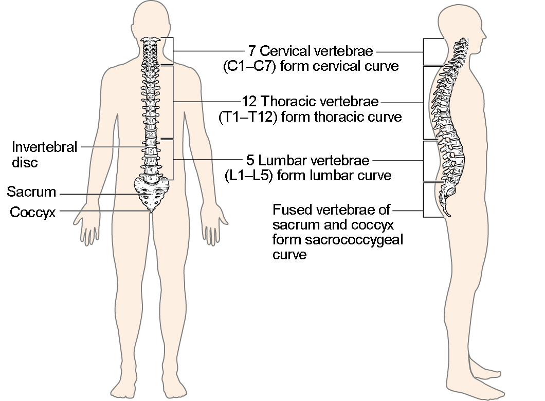
The Vertebral Column Anatomy And Physiology I from s3-us-west-2.amazonaws.com The vertebral column is a series of approximately 33 bones called vertebrae, which are separated by intervertebral discs. Carpals—8 small bones of the wrist — includes the scaphoid, lunate, capitate, trapezium, and others. As a person ages, these bones grow together and fuse into larger bones, leaving adults with only 206 bones. It also covers some common conditions and injuries that can affect the back. Human backbone diagram, bone, human backbone diagram. The human skeleton, like that of other vertebrates, consists of two principal subdivisions, each with origins distinct from the others and each presenting certain individual features.these are (1) the axial, comprising the vertebral column—the spine—and much of the skull, and (2) the appendicular, to which the pelvic (hip) and pectoral (shoulder) girdles and the bones and cartilages of the. Cervical bones diagram 12 photos of the cervical bones diagram cervical bones diagram, cervix anatomy diagram related posts of human back bones diagram human bone parts name. Muscles of the abdomen lower back and pelvis.
They help support particular bones and make them move.
Pelvic bone labeled 12 photos of the pelvic bone labeled pelvic bone labeled, pelvic bone labeling quiz, pelvic bone with labeling, pelvic girdle bone labeling quiz, pubic bone labeled, bone, pelvic bone labeled, pelvic bone labeling quiz, pelvic bone with labeling, pelvic girdle bone labeling quiz, pubic bone labeled Spinal vertebrae bone spine vertebra toracica spinal cord spine structure back diagram spine sections spinal cord vertebrae spinal structure health diagram. The human skeleton, like that of other vertebrates, consists of two principal subdivisions, each with origins distinct from the others and each presenting certain individual features.these are (1) the axial, comprising the vertebral column—the spine—and much of the skull, and (2) the appendicular, to which the pelvic (hip) and pectoral (shoulder) girdles and the bones and cartilages of the. Label the bones on the skeleton. The vertebral column houses the spinal canal, a cavity that. For more anatomy content please follow us and visit our website: Our latest youtube film is ready to run. Bones of the pelvis and lower back. Related posts of female body back side anatomy skeleton bones diagram. As a person ages, these bones grow together and fuse into larger bones, leaving adults with only 206 bones. Muscles of the abdomen lower back and pelvis. Cheek bone (zygoma) upper jaw (maxilla). Anatomical diagrams of the spine and back.
They are the bones of your forearm human back bones. The smallest bone in the human body is called the stirrup bone, located deep inside the ear.